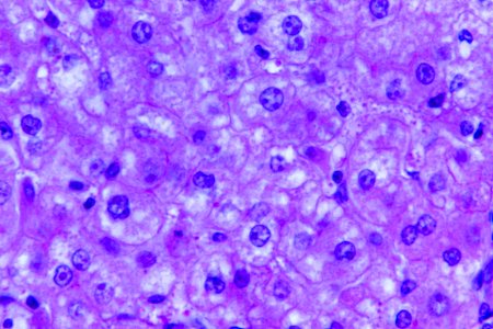
A 440X photomicrograph magnification of a hematoxylin and eosin (H&E)–stained liver tissue specimen, revealed the presence of cytoarchitectural changes indicative of fatty degeneration. Original image sourced from US Government department: Public Health Image Library, Centers for Disease Control and Prevention. Under US law this image is copyright free, please credit the government department whenever you can”.